Antimony »
PDB 1exi-8cgs »
6xlk »
Antimony in PDB 6xlk: Cryo-Em Structure of Ecmrr-Dna Complex in Ecmrr-Rpitc-4NT
Antimony Binding Sites:
The binding sites of Antimony atom in the Cryo-Em Structure of Ecmrr-Dna Complex in Ecmrr-Rpitc-4NT
(pdb code 6xlk). This binding sites where shown within
5.0 Angstroms radius around Antimony atom.
In total 2 binding sites of Antimony where determined in the Cryo-Em Structure of Ecmrr-Dna Complex in Ecmrr-Rpitc-4NT, PDB code: 6xlk:
Jump to Antimony binding site number: 1; 2;
In total 2 binding sites of Antimony where determined in the Cryo-Em Structure of Ecmrr-Dna Complex in Ecmrr-Rpitc-4NT, PDB code: 6xlk:
Jump to Antimony binding site number: 1; 2;
Antimony binding site 1 out of 2 in 6xlk
Go back to
Antimony binding site 1 out
of 2 in the Cryo-Em Structure of Ecmrr-Dna Complex in Ecmrr-Rpitc-4NT
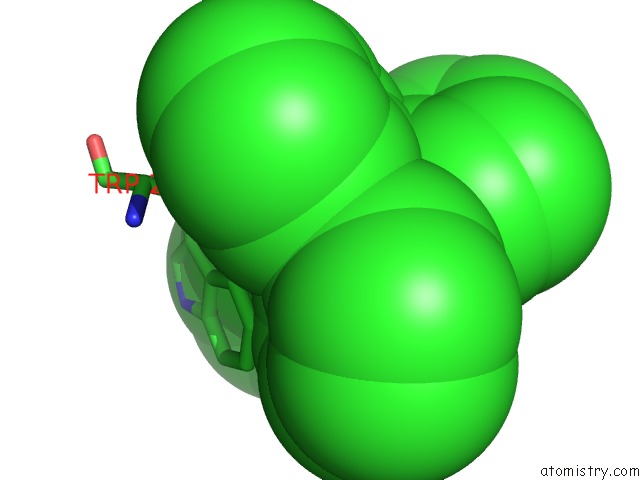
Mono view
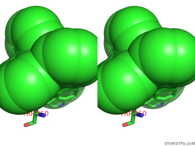
Stereo pair view
Mono view
Stereo pair view
A full contact list of Antimony with other atoms in the Sb binding
site number 1 of Cryo-Em Structure of Ecmrr-Dna Complex in Ecmrr-Rpitc-4NT within 5.0Å range:
|
Antimony binding site 2 out of 2 in 6xlk
Go back to
Antimony binding site 2 out
of 2 in the Cryo-Em Structure of Ecmrr-Dna Complex in Ecmrr-Rpitc-4NT
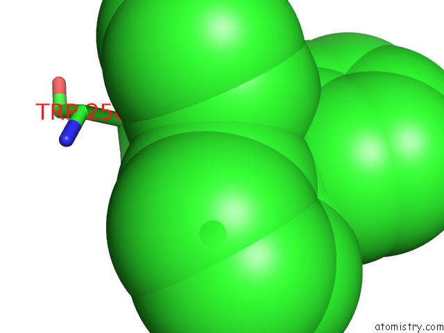
Mono view
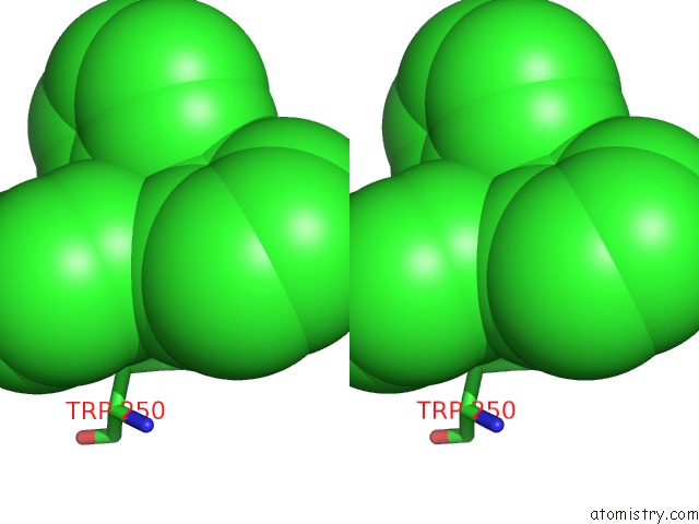
Stereo pair view
Mono view
Stereo pair view
A full contact list of Antimony with other atoms in the Sb binding
site number 2 of Cryo-Em Structure of Ecmrr-Dna Complex in Ecmrr-Rpitc-4NT within 5.0Å range:
|
Reference:
Y.Yang,
C.Liu,
W.Shi,
D.G.Schatz,
Y.Hu,
B.Liu.
Structural Visualization of Bacterial Multidrug-Activated Transcription To Be Published.
Page generated: Thu Oct 10 13:18:34 2024
Last articles
Zn in 9MJ5Zn in 9HNW
Zn in 9G0L
Zn in 9FNE
Zn in 9DZN
Zn in 9E0I
Zn in 9D32
Zn in 9DAK
Zn in 8ZXC
Zn in 8ZUF