Antimony »
PDB 1exi-8cgs »
1f48 »
Antimony in PDB 1f48: Crystal Structure of the Escherichia Coli Arsenite-Translocating Atpase
Protein crystallography data
The structure of Crystal Structure of the Escherichia Coli Arsenite-Translocating Atpase, PDB code: 1f48
was solved by
T.Zhou,
S.Radaev,
B.P.Rosen,
D.L.Gatti,
with X-Ray Crystallography technique. A brief refinement statistics is given in the table below:
| Resolution Low / High (Å) | 26.37 / 2.30 |
| Space group | I 2 2 2 |
| Cell size a, b, c (Å), α, β, γ (°) | 73.523, 75.715, 222.714, 90.00, 90.00, 90.00 |
| R / Rfree (%) | 20.6 / 26.3 |
Other elements in 1f48:
The structure of Crystal Structure of the Escherichia Coli Arsenite-Translocating Atpase also contains other interesting chemical elements:
| Cadmium | (Cd) | 6 atoms |
| Magnesium | (Mg) | 2 atoms |
| Chlorine | (Cl) | 3 atoms |
Antimony Binding Sites:
The binding sites of Antimony atom in the Crystal Structure of the Escherichia Coli Arsenite-Translocating Atpase
(pdb code 1f48). This binding sites where shown within
5.0 Angstroms radius around Antimony atom.
In total 4 binding sites of Antimony where determined in the Crystal Structure of the Escherichia Coli Arsenite-Translocating Atpase, PDB code: 1f48:
Jump to Antimony binding site number: 1; 2; 3; 4;
In total 4 binding sites of Antimony where determined in the Crystal Structure of the Escherichia Coli Arsenite-Translocating Atpase, PDB code: 1f48:
Jump to Antimony binding site number: 1; 2; 3; 4;
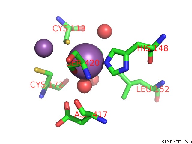
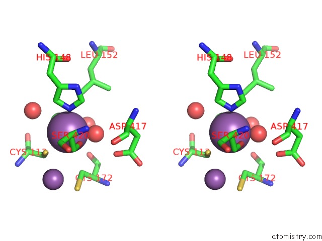
Antimony binding site 1 out of 4 in 1f48
Go back to
Antimony binding site 1 out
of 4 in the Crystal Structure of the Escherichia Coli Arsenite-Translocating Atpase
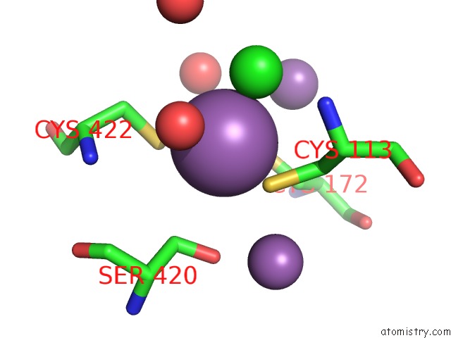
Mono view
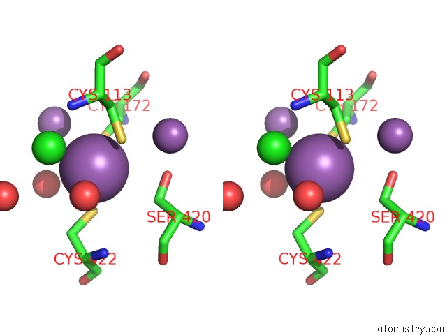
Stereo pair view
Mono view
Stereo pair view
A full contact list of Antimony with other atoms in the Sb binding
site number 1 of Crystal Structure of the Escherichia Coli Arsenite-Translocating Atpase within 5.0Å range:
|
Antimony binding site 2 out of 4 in 1f48
Go back to
Antimony binding site 2 out
of 4 in the Crystal Structure of the Escherichia Coli Arsenite-Translocating Atpase
Mono view
Stereo pair view
Mono view
Stereo pair view
A full contact list of Antimony with other atoms in the Sb binding
site number 2 of Crystal Structure of the Escherichia Coli Arsenite-Translocating Atpase within 5.0Å range:
|
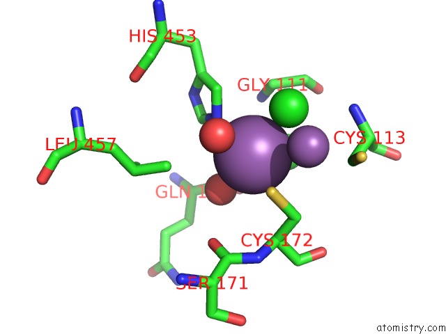
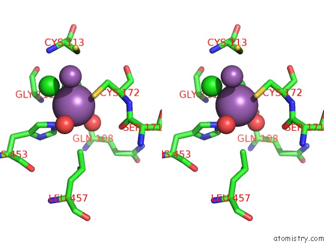
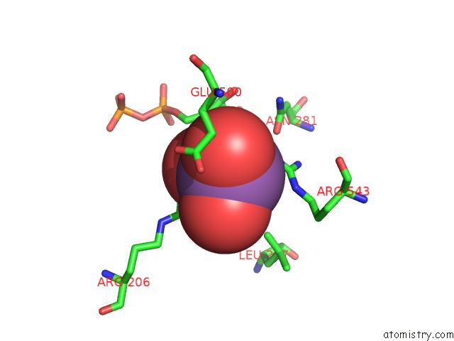
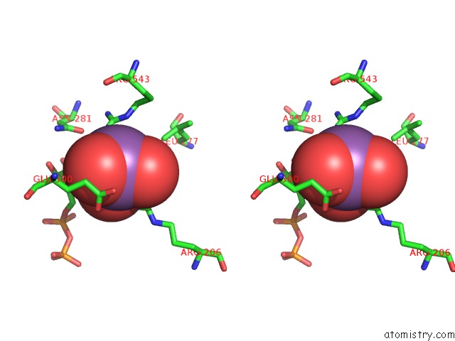
Antimony binding site 3 out of 4 in 1f48
Go back to
Antimony binding site 3 out
of 4 in the Crystal Structure of the Escherichia Coli Arsenite-Translocating Atpase
Mono view
Stereo pair view
Mono view
Stereo pair view
A full contact list of Antimony with other atoms in the Sb binding
site number 3 of Crystal Structure of the Escherichia Coli Arsenite-Translocating Atpase within 5.0Å range:
|
Antimony binding site 4 out of 4 in 1f48
Go back to
Antimony binding site 4 out
of 4 in the Crystal Structure of the Escherichia Coli Arsenite-Translocating Atpase
Mono view
Stereo pair view
Mono view
Stereo pair view
A full contact list of Antimony with other atoms in the Sb binding
site number 4 of Crystal Structure of the Escherichia Coli Arsenite-Translocating Atpase within 5.0Å range:
|
Reference:
T.Zhou,
S.Radaev,
B.P.Rosen,
D.L.Gatti.
Structure of the Arsa Atpase: the Catalytic Subunit of A Heavy Metal Resistance Pump. Embo J. V. 19 4838 2000.
ISSN: ISSN 0261-4189
PubMed: 10970874
DOI: 10.1093/EMBOJ/19.17.4838
Page generated: Thu Oct 10 13:17:38 2024
ISSN: ISSN 0261-4189
PubMed: 10970874
DOI: 10.1093/EMBOJ/19.17.4838
Last articles
Cl in 6AHYCl in 6AHH
Cl in 6AH9
Cl in 6AGR
Cl in 6AGN
Cl in 6AFJ
Cl in 6AFL
Cl in 6AFI
Cl in 6AFH
Cl in 6AFF